Santiago Ramón y Cajal (1852-1934)
Santiago Ramón y Cajal was awarded with the Nobel Prize in Physiology or Medicine in 1906 and in 1907 he was appointed as Chair of the Board of the Junta para Ampliación de Estudios e Investigaciones Biológicas (JAE, 1907-1939), which was part of the Spanish Ministry of Public Instruction and Fine Arts.
As JAE's President (1907-1932), Cajal led the largest scientific regeneration and modernization project carried out in Spain in the early 20th century. During his long presidency, he encouraged structural changes in the Spanish educational system. Cajal’s JAE was the origin of the Spanish National Research Council (CSIC).
In 1902, Cajal was appointed director of the Laboratorio de Investigaciones Biológicas, a research centre founded by order of HM the King Alfonso XIII to distinguish the Moscow Prize awarded to Santiago Ramón y Cajal in 1900 at the International Congress of Medicine held in Paris. This national laboratory gave rise to the Cajal Institute in 1922, which was then incorporated to the CSIC the 24th November 1939.
Santiago Ramón y Cajal is often called “the father of modern neuroscience” for his outstanding studies on the microscopic anatomy of the nervous system, his observations regarding its degeneration and regeneration, and his theories about the function, development and plasticity of virtually the whole central nervous system. For the first time, Cajal placed Spain in the forefront of international science. Cajal was awarded with the Nobel Prize in 1906. After all this time, his almost fifty years of work (1887-1934) continue to captivate and stimulate modern neuroscientists worldwide. In 1888 (Figures 1, 2), he discovered that the nervous system, including the brain, is composed of individual entities, later termed neurons.
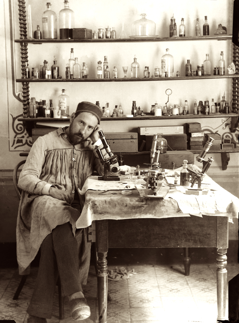

Figures 1, 2 Self-portraits taken by Cajal in his laboratory in Valencia (Spain) during his early thirties, c. 1885-1887.
His findings disproved the popular reticular theory, which viewed the nervous system as a continuous network of fibers (Figure 3).
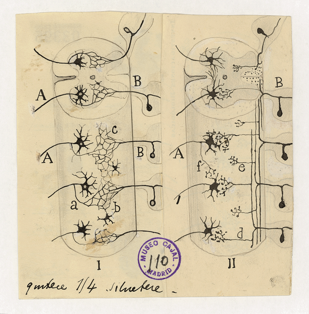
Figure 3.- Cajal’s drawing explaining the differences between the neuron and reticular theories: “Scheme to compare the concept of Golgi regarding the sensory- motor connections of the spinal cord (I) with the results of my researchs (II). A, anterior roots; B, posterior roots; a, collateral of a motor root; b, cells with a short axon which, according to Golgi, would intervene in the formation of the network; c, diffuse interstitial network; d, our long collaterals in contact with the motor cells; e, short collaterals.” This figure was reproduced as Fig. 9 in Recuerdos de mi vida. Historia de mi labor científica (Recollections of my life — The story of my scientific work”. Original drawing by Santiago Ramón y Cajal, black Chinese ink on paper, c. 1923.
Cajal studied every phase in the lives of neurons. In embryos, he observed a dynamic structure at the tip of developing axons (the growth cone), which he hypothesized might be guided by chemicals (neurotropism) (Figure 4).
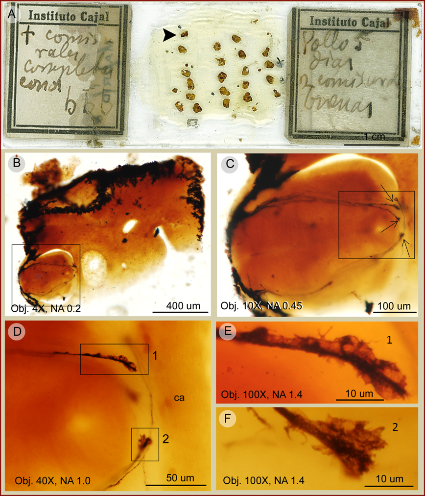
Figure 4.- Growth cones detected in a Cajal´s histological preparation of the spinal cord of a day 5 chick embryo. The slide is preserved at the Instituto Cajal. Panel A, shows the aspect of a Cajal´s original slide impregnated by the Golgi method. Notice the label handwritten by Cajal stating: "† comisurales completas conos bbb" ["† complete commissural cones bbb"; "b" means “bien" in Spanish, "good" in English], "Pollo 5 días 2 comisurales buenas" ("Chick 5 days 2 good commissurals"). Panel B, illustrates details of a section in A (arrowhead). Panel C, depicts a higher-power magnifications of the squared area in B. Notice in C a number of growth cones (some pointed by arrows). Panels D-F, show details of the growth cones shown in C. Notice the excellent preservation of the sample.
He inferred that, because of the spaces between them, neurons must communicate not by continuity but by contact (later termed synapse) (Figure 5).
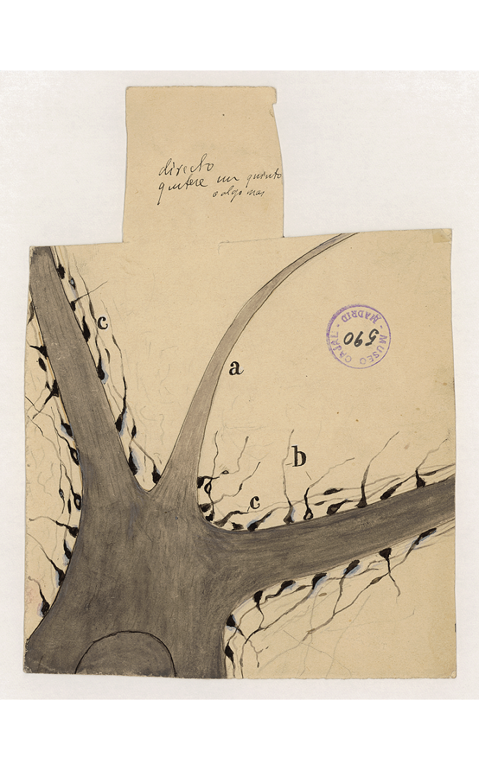
Figure 5.- Motoneuron with its axon (a) and dendrites (b) and synaptic terminal buttons (c) on it. Partial view. Original drawing by Santiago Ramón y Cajal, black Chinese ink on paper, c. 1909.
From static images alone, he was able to determine the general flow of nervous activity (the law of dynamic polarization) (Figure 6).
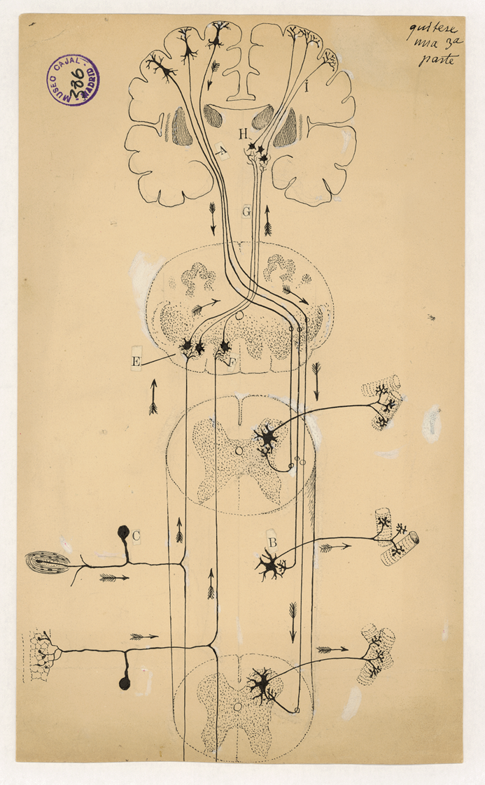
Figure 6.- Schematic drawing of the motor and sensitive pathways. Original drawing by Santiago Ramón y Cajal, black Chinese ink on paper, c. 1899.
Cajal also identified that there are protrusions on the stems of dendrites (dendritic spines), which his contemporaries dismissed as artifacts but which he recognized as vital extra contact sites (Figure 7).
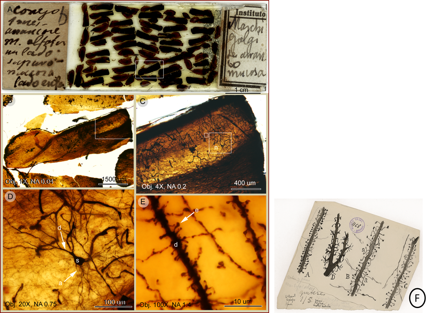
Figura 7.- Dendritic spines detected in a Cajal´s histological preparation through the rabbit olfactory bulb. The slide is preserved at the Instituto Cajal. Panel A shows the aspect of a Cajal´s original slide impregnated by the Golgi/ Marchi's method. Notice the labels handwritted by Cajal: left hand side, stating the animal species: “Conejo 1 mes” — “1-month old rabbit” —, and on the right hand side the staining procedure “Marchi Golgi”. Panels B and C illustrate details of a section in A (squared). Panel D depicts a neuron with its components: s, soma; d, dendrite & a, axon. Panel E represents a higher power magnification of the dendrite in D. Notice in panel E numerous dendritic spines (e) along the same dendrite shown in D. Also note the excellent preservation of the sample. Dendritic spines were accurately drawn by Cajal: Panel F scientific drawing of Santiago Ramón y Cajal, representing types of collateral spines of cerebral pyramids. Original drawing by Santiago Ramón y Cajal, black Chinese ink on paper, c. 1899.
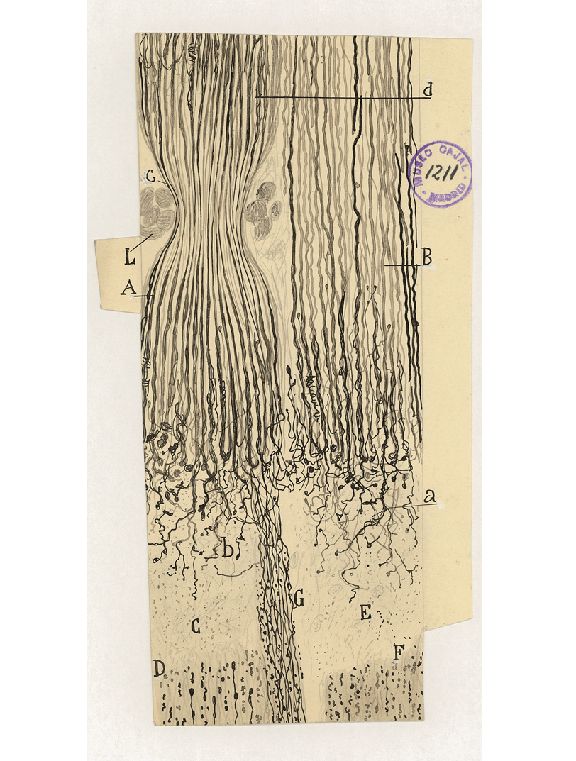
In the 1890s, Cajal was among the first to comment on the capacity of neurons to adapt their morphology (plasticity) (Figure 8). In fact, he may have been responsible for popularising the term: “If he is so determined,” Cajal said, “man can become the sculptor of his own brain.”
Figura 8.- Nearly 100 years ago, Ramon y Cajal used the term “abortive regeneration” to describe the attempted regrowth of injured neurons in a model of sciatic nerve injury in young cats and rabbits killed few days after ligature. Results on regeneration and degeneration of the nervous system were accurately drawn by Cajal. As an example, this panel represents semi-ligature of the sciatic nerve in a rabbit killed eight days after the operation. Total section of the nerve, near the ligature, is shown in order to compare the sprouting capacity of the two portions. A, peripheral stump of the ligated nerve. B, central stump of the non-ligated fascicle. C, E, scars. d, central stump. L, ligature. D, F, degenerated stumps. Original drawing by Santiago Ramón y Cajal, black Chinese ink on paper, c. 1899.
Cajal’s masterpiece, The Histology of the Nervous System of Man and Vertebrates, is still quoted hundreds of times each year. His Degeneration and Regeneration of the Nervous System and The Structure of the Retina publications are considered classics. During his career, Cajal published over three hundred articles, not all of which were neuroscientific. It is a little known fact that he discovered the cholera vaccine. He also contributed significantly to the study of cancer. In addition, Cajal was a pioneer of colour photography (La fotografía de los colores). He published short fiction (Vacation Stories), a collection of worldly wisdom (Charlas de café), an account of extreme old age (El mundo visto a los ochenta años), a scientific guidebook (Advice for a Young Researcher) and an unforgettable autobiography (Recollections of My Life). When Cajal won the 1906 Nobel Prize in 1906, he became a national hero. To this day, there is a street named after him in virtually every Spanish city.
Significance of the CSIC’s “Cajal Legacy”
Most of the original possessions of Cajal are housed at the Cajal Institute in Madrid, which belongs to the CSIC, at Avenida Doctor Arce. Only a few objects of this patrimony are permanently exhibited at the Institute’s library, where the workplace of Cajal is recreated.
The Cajal Legacy was inventoried in 2008. It includes 28.222 items, that can be catalogued and listed as follows:
- 1. Photographic archive (2.773)
- 2. Precision balances (2)
- 3. Photographic cameras (5)
- 4. Correspondence (2.584)
- 5. Ceramics (2)
- 6. Colorants, Reagents and Solutions (387)
- 7. Notebooks (11)
- 8. Scientific drawings (1.976), from which 1800 belong to Cajal and other members of his school
- 9. Artistic drawings (2)
- 10. Diplomas and Certificates (109)
- 11. Sculptures (6)
- 12. Phonographs (1)
- 13 Books, Newspapers and Journals (7.000)
- 14. Handwritten manuscripts (1.952)
- 15. Gas lighters (3)
- 16. Medals, Decorations and Awards (25), including the gold medal of von Helmholtz, the Nobel Prize and the Echegaray award
- 17. Optic microscopes (21), Boxes (5) and Microphotography devices (1)
- 18. Microtomes (3)
- 19. Furniture (20), among which are included his work table and chair, cabinets for storing chemical products and vitrines.
- 20. Straight razors (9)
- 21. Personal objects (15) such as his last glasses, wallet, walking stick, passport, identity document, toga and camera.
- 22. Paintings (10)
- 23. Histological slides (17.150, from which 3.000 are originals by Cajal)
- 24. Projectors (4)
- 25. Telescope (1), employed in Cajal’s researches
- 26. Textiles
In thus Legacy are also included the items produces by the scientific work of the disciples of Cajal (Escuela de Histología Española de Cajal).
For more details visit: https://www.ncbi.nlm.nih.gov/pmc/articles/PMC2845060/
Value of the drawings as a graphical illustration of the microscopic observations
In Cajal's time, scientists generally used drawing as the means to illustrate microscopic observations. Therefore, accepting published findings was often an act of faith. Revolutionary histological drawings by Cajal were initially considered by some researchers as artistic interpretations rather than accurate copies of his preparations. But Cajal's drawings are pieces of reality, reliable copies of histological preparations that show the microorganisation of the nervous system: the delicate structure of brain cells and their connections. While his contributions to current concepts of brain function and organisation are widely publicised, his carefully executed drawings — displaying a rare mix of artistic skill and scientific insight — are much less known. Hundreds of his drawings are housed in the Cajal Legacy. Cajal’s histological drawings are not only valuable for their beauty but also because they express universal concepts and are still used for educational and training purposes today.
Virtual visit of the Cajal Legacy: http://www.cajal.csic.es/legado.html